Percutaneous vertebroplasty
1. The patient was placed in prone position and disinfected locally. Local anesthesia or general anesthesia were used as appropriate.
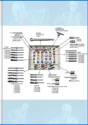
2. Puncture needle should be selected according to the need. The pedicle approach is usually used to puncture under fluoroscopy, and the core of puncture needle should be taken out after puncture is in place.
3. Inhale the prepared bone cement (PMMA) with a special syringe and install the screw propeller to fix it firmly.
4. Rotating the push rod of the screw propeller to observe the state of the extruded bone cement. When toothpaste is formed, the syringe is quickly connected to the needle and injected under fluoroscopy to observe the distribution of bone cement in the vertebral body.
5. After the injection, remove the screw propeller, insert the core of the puncture needle, pull out the puncture needle, bandage locally, and finish the operation.
Percutaneous kyphoplasty
1. Under fluoroscopy, the dismountable needle pedicle puncture needle enters the collapsed vertebral body through the pedicle. After confirmation, the needle seat is removed, the orthotic sleeve is inserted along the needle core, and then the needle core is taken out to establish a working channel.
2. Insert the vertebral drill into the orthopedic sleeve (if biopsy is needed, hollow vertebral drill can be used). Observe under fluoroscopy and remove the vertebral drill after reaching the required depth.
3. The balloon was put into the collapsed vertebral body and fluoroscopy confirmed that the balloon should all extend the
orthopedic sleeve (two marking rings of the head of the balloon can be seen).
4. The balloon was dilated by slowly injecting contrast agent under fluoroscopy with a pressure gauge (attached to the method of use). The collapsed vertebral body was elevated to form a cavity, the pressure and volume were measured, the contrast agent was sucked out, and the balloon was withdrawn slowly after retracting to vacuum.
4. Under fluoroscopy, cement was injected into the vertebral body, then the working cannula was pulled out and the operation was completed.
 +86-021-50327060
 zq@lzqtech.com
 Medical Instrument
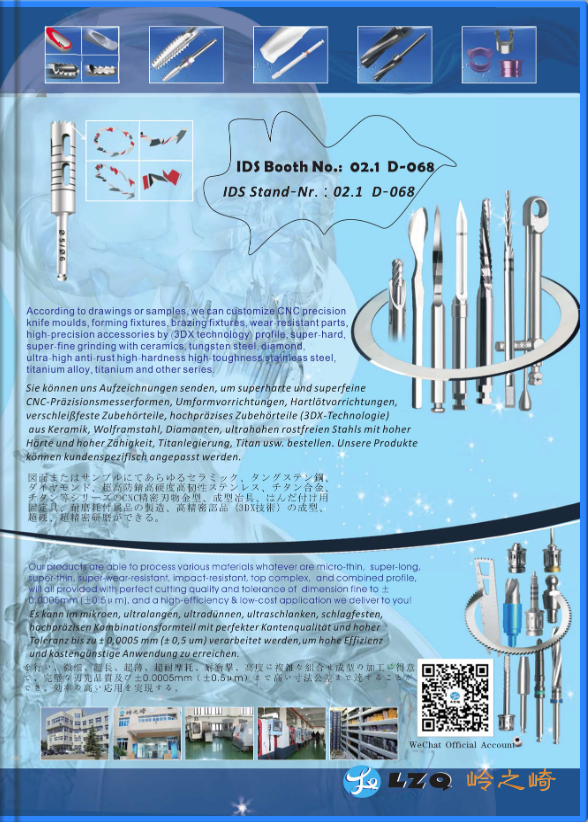
We can achieve perfect edge quality and dimensional tolerance up to±0.0005mm (±0.5μm) in the process of micro, ultra-long, ultra-thin, super-abrasive, impact-resistant, high-precision and combined ... VIEW MORE
Medical Instrument
We can achieve perfect edge quality and dimensional tolerance up to±0.0005mm (±0.5μm) in the process of micro, ultra-long, ultra-thin, super-abrasive, impact-resistant, high-precision and combined ... VIEW MORE Implant
Corresponding and matching drills and tools of different types, forms, shapes, structures can be high precisely ground to mold according to different brands and different types of implants forms, shap... VIEW MORE
Implant
Corresponding and matching drills and tools of different types, forms, shapes, structures can be high precisely ground to mold according to different brands and different types of implants forms, shap... VIEW MORE Cutting Tools
Super-hardness machining for special, non-standard new ite ms with different size and tolerance. VIEW MORE
Cutting Tools
Super-hardness machining for special, non-standard new ite ms with different size and tolerance. VIEW MORE Accessory Parts
we can customize for you according to your samples or drawings for any manufacturing of ceramic,carbide,stainless high-speed steel, stainless steel, titanium alloy, titanium diamond, etc series, hig... VIEW MORE
Accessory Parts
we can customize for you according to your samples or drawings for any manufacturing of ceramic,carbide,stainless high-speed steel, stainless steel, titanium alloy, titanium diamond, etc series, hig... VIEW MORE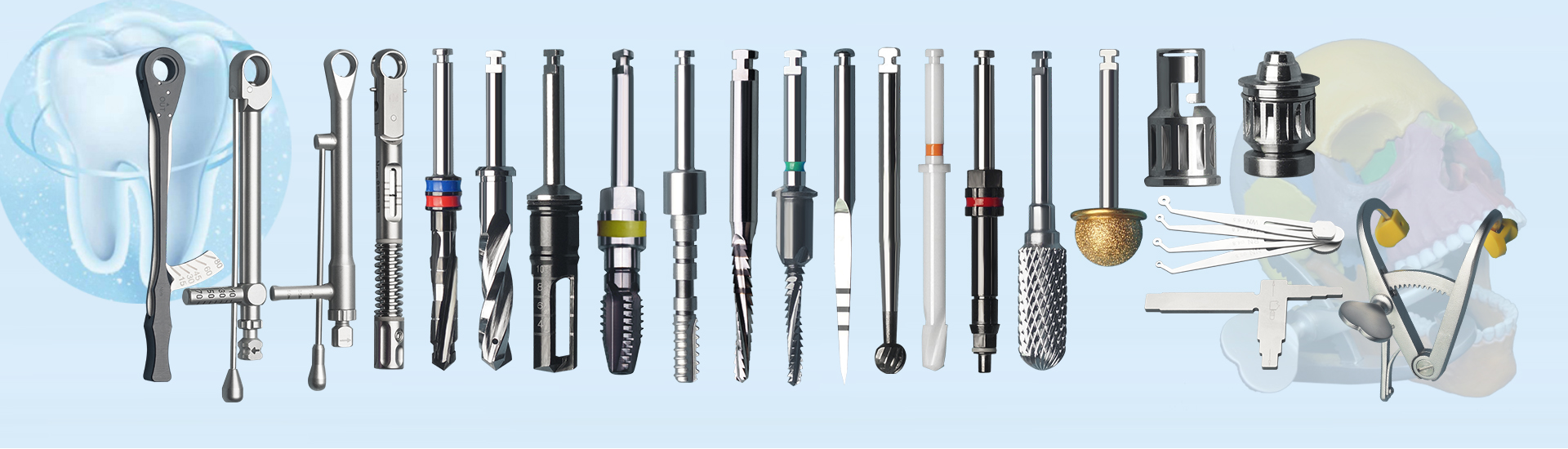
 +86-021-50327060
 NO.1269 Plant, Jinhu Road, Jinqiao Export Processing Zone, Pudong New District, Shanghai, China.